Cranial Nerve Easy to Remember Drawing
Creating a Simple Drawing of Cranial Nerve Pathways

Cranial nerve easy to remember drawing – Okay, so you wanna draw some cranial nerves? Think of it like a super-powered, miniature highway system for your brain. We’re going to keep it simple, like a back-of-the-napkin sketch that’ll still impress your neuroanatomy professor (or your friends, who totally
need* to know this stuff).
Creating memorable diagrams for cranial nerves often involves simplifying complex structures. A helpful mnemonic technique might involve visualizing the nerves’ pathways as beams of light, similar to the straightforward approach shown in this tutorial on sunlight drawing beaming down easy drawing. This analogy aids in understanding the directional flow and spatial relationships of these crucial nerves, ultimately improving memorization of their functions and locations.
This diagram will show the brain and the pathways of each cranial nerve, using arrows to show where the signals are going. We’ll also number each nerve and pinpoint its nucleus location within the brainstem. Think of it as a neural network map for your head – seriously cool stuff.
Cranial Nerve Pathway Diagram
Imagine a simple oval representing the brain. From this oval, draw twelve pairs of lines extending outwards, representing the twelve cranial nerves. These lines should be slightly curved to mimic the actual pathways. Each line should have an arrow indicating the direction of nerve impulse transmission – either towards or away from the brain. For sensory nerves, the arrow points towards the brain; for motor nerves, it points away.
Mixed nerves will have arrows pointing both ways.The brainstem, located at the base of the brain, should be represented as a slightly thicker, irregular shape connected to the oval. Within the brainstem, small dots or circles can represent the nuclei of each cranial nerve. These nuclei are grouped regionally, and their positions will be described in the following section.
Don’t worry about making it anatomically perfect; the goal is clarity and understanding, not a medical illustration for a textbook. Think of it like drawing a simplified subway map – you get the gist, even if it’s not totally to scale.
Cranial Nerve Nuclei Locations
Now, let’s pinpoint those nuclei within our brainstem. This part’s like playing a game of “Where’s Waldo?”, but with brain structures. The locations are approximate and simplified for this basic diagram. Remember, the brainstem is comprised of the midbrain, pons, and medulla oblongata.
- Olfactory Nerve (CN I): This one’s a bit of an outlier; its receptor cells are in the nasal cavity, and it doesn’t directly originate from the brainstem. You can simply draw it entering the brain near the frontal lobe.
- Optic Nerve (CN II): Similar to CN I, its receptors are in the retina. Show it entering the brain near the diencephalon.
- Oculomotor Nerve (CN III), Trochlear Nerve (CN IV), Abducens Nerve (CN VI): These nerves control eye movement. Their nuclei are located in the midbrain. Draw their nuclei clustered together in this region of your brainstem.
- Trigeminal Nerve (CN V): This nerve has sensory and motor functions for the face. Its nuclei are spread across the pons and midbrain. Indicate this by showing multiple nuclei for this nerve within those regions.
- Abducens Nerve (CN VI): As mentioned above, it controls eye movement and its nucleus is in the pons.
- Facial Nerve (CN VII): Controls facial expression and taste. Its nuclei are primarily located in the pons.
- Vestibulocochlear Nerve (CN VIII): Deals with hearing and balance. Its nuclei are in the pons and medulla.
- Glossopharyngeal Nerve (CN IX), Vagus Nerve (CN X), Accessory Nerve (CN XI): These nerves are involved in swallowing, speech, and parasympathetic functions. Their nuclei are clustered in the medulla.
- Hypoglossal Nerve (CN XII): Controls tongue movement. Its nucleus is located in the medulla.
Remember, this is a simplified representation. The actual pathways and nuclear locations are far more complex. But this basic diagram should provide a solid foundation for understanding the general layout of the cranial nerves. Now go forth and draw!
Illustrating Cranial Nerve Functions
Okay, so you’ve got your basic cranial nerve pathways down – awesome! Now let’s crank it up a notch and visualize what each of these nerves
actually* does. Think of it like this
each nerve is a superstar with a specific role in the grand, amazing production that is your body. We’re going to give each one its own spotlight moment.
Understanding cranial nerve function isn’t just about memorizing names and numbers; it’s about seeing how they work together to create the symphony of sensation and movement that defines us. Damage to any one nerve can throw off the whole performance, so let’s get to know the players.
Cranial Nerve Functions and Damage Consequences
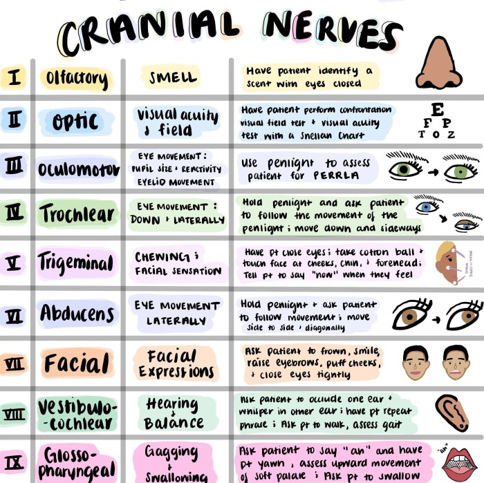
The following table provides a simplified overview of the twelve cranial nerves, their functions, and the potential consequences of damage. Think of this as your backstage pass to the nervous system’s biggest show!
| Nerve Number | Nerve Name | Function (Simplified) | Illustration & Damage Consequences |
|---|---|---|---|
| I | Olfactory | Smell | A nose sniffing a flower. Damage: Anosmia (loss of smell). Imagine never smelling your favorite pizza again – bummer! |
| II | Optic | Vision | An eye looking at a vibrant scene. Damage: Visual field loss, blindness. Picture missing that amazing sunset… tragic. |
| III | Oculomotor | Eye movement, pupil constriction | An eye moving in different directions. Damage: Double vision (diplopia), drooping eyelid (ptosis), dilated pupil. Think blurry vision and a perpetually surprised look. |
| IV | Trochlear | Eye movement (superior oblique muscle) | An eye looking downward and inward. Damage: Difficulty looking downward and inward. Like trying to read a book held really low – annoying! |
| V | Trigeminal | Facial sensation, chewing | A face with marked areas for sensation and jaw movement. Damage: Facial numbness, weakness in chewing muscles. Think difficulty feeling a touch on your face, or struggling to bite into a juicy steak. |
| VI | Abducens | Eye movement (lateral rectus muscle) | An eye looking laterally (to the side). Damage: Inability to move eye laterally. Like having a blind spot that follows you around. |
| VII | Facial | Facial expression, taste (anterior 2/3 of tongue) | A smiling face. Damage: Facial paralysis (Bell’s palsy), loss of taste. Imagine trying to smile for a selfie and only one side of your face moves… awkward! |
| VIII | Vestibulocochlear | Hearing, balance | An ear and a representation of balance. Damage: Hearing loss (deafness), vertigo (dizziness), imbalance. Picture constantly feeling like the room is spinning – not fun! |
| IX | Glossopharyngeal | Taste (posterior 1/3 of tongue), swallowing, salivation | The back of the tongue and throat. Damage: Difficulty swallowing, loss of taste, decreased salivation. Think choking on your food and a super dry mouth. |
| X | Vagus | Parasympathetic control of many organs | A schematic of the body showing the vagus nerve’s widespread influence. Damage: Difficulty swallowing, hoarseness, changes in heart rate and digestion. A whole host of problems! |
| XI | Accessory | Shoulder and neck movement | Shoulders and neck muscles. Damage: Weakness in shoulder and neck muscles. Think struggling to lift a grocery bag or turn your head. |
| XII | Hypoglossal | Tongue movement | A tongue sticking out. Damage: Difficulty with speech (dysarthria), swallowing. Imagine slurring your words and struggling to eat. |
Clinical Correlation: Cranial Nerve Easy To Remember Drawing

Think of this section as your cheat sheet for acing a cranial nerve exam – or, you know, for actually helping patients. We’re going to break down how doctors test each cranial nerve and what happens when things go wrong. It’s like a detective story, but instead of solving crimes, we’re solving neurological mysteries!
Cranial Nerve I: Olfactory Nerve Examination
Testing the olfactory nerve is all about smell. Doctors typically use familiar, non-irritating scents like coffee, peppermint, or cloves. They’ll have the patient close one nostril at a time and identify the smell. Damage to the olfactory nerve can result in anosmia (loss of smell), which can be caused by anything from a stuffy nose to a head injury.
Imagine not being able to smell your favorite pizza – bummer!
Cranial Nerve II: Optic Nerve Examination
Visual acuity is key here. Doctors will use a Snellen chart (that big eye chart you see in the doctor’s office) to assess visual sharpness. They’ll also check visual fields (peripheral vision) using confrontation testing – basically, comparing the patient’s vision to their own. Damage to the optic nerve can lead to visual field defects (like blind spots) or even complete vision loss, depending on the severity and location of the damage.
Think of it like a glitch in your visual matrix.
Cranial Nerves III, IV, and VI: Oculomotor, Trochlear, and Abducens Nerve Examination
These three nerves control eye movements. Doctors assess pupil size and reactivity to light (checking for things like unequal pupil size or sluggish responses), and they test eye movements in all directions. They’ll follow a finger or penlight with their eyes. Damage can cause things like double vision (diplopia), drooping eyelids (ptosis), or an inability to move the eyes in certain directions.
Imagine trying to play a game of Pong with your eyes, but your eyes are totally out of sync.
Cranial Nerve V: Trigeminal Nerve Examination
The trigeminal nerve has three branches that control sensation in the face and chewing. Doctors will test sensation (light touch, pain, temperature) on the face using various tools and ask the patient to clench their jaw to assess motor function. Damage can lead to facial numbness, pain (like trigeminal neuralgia, which is excruciating), or weakness in the jaw muscles.
Think of it as a malfunction in your face’s personal sensory system.
Cranial Nerve VII: Facial Nerve Examination
This nerve controls facial expressions and taste. Doctors assess symmetry of facial movements (smile, frown, raise eyebrows) and test taste on the anterior two-thirds of the tongue. Damage can result in facial paralysis (Bell’s palsy is a common example), affecting the ability to smile, frown, or close one eye properly. It’s like your face forgot how to do its job.
Cranial Nerve VIII: Vestibulocochlear Nerve Examination
Hearing and balance are the stars here. Doctors test hearing acuity with a whisper test or tuning fork tests (like the Rinne and Weber tests). They also assess balance by checking for gait abnormalities or nystagmus (involuntary eye movements). Damage can cause hearing loss (conductive or sensorineural), tinnitus (ringing in the ears), or vertigo (dizziness). Imagine a world where your balance is off and your ears are constantly ringing – a recipe for disaster!
Cranial Nerve IX: Glossopharyngeal Nerve Examination, Cranial nerve easy to remember drawing
This nerve involves swallowing, taste, and sensation in the back of the throat. Doctors will check the gag reflex, assess taste on the posterior third of the tongue, and evaluate swallowing ability. Damage can cause difficulty swallowing (dysphagia), loss of taste, or a diminished gag reflex. Imagine choking on your food every time you try to eat – not fun!
Cranial Nerve X: Vagus Nerve Examination
The vagus nerve controls many functions, including swallowing, voice production, and parasympathetic activity. Doctors assess swallowing, hoarseness, and the gag reflex. Damage can cause hoarseness, difficulty swallowing, or changes in heart rate or blood pressure. Think of it as the maestro of your internal orchestra, and when it’s off, things get chaotic.
Cranial Nerve XI: Accessory Nerve Examination
This nerve controls the sternocleidomastoid and trapezius muscles, responsible for head turning and shoulder shrugging. Doctors will ask the patient to turn their head against resistance and shrug their shoulders. Damage can lead to weakness in these muscles, making it difficult to turn the head or lift the shoulders. Imagine struggling to hold up your own head or carry a heavy bag – tough times.
Cranial Nerve XII: Hypoglossal Nerve Examination
This nerve controls tongue movement. Doctors will ask the patient to stick out their tongue and move it side to side. Damage can cause tongue weakness or deviation (the tongue will drift to one side). Imagine trying to lick an ice cream cone, but your tongue just won’t cooperate.
FAQ Explained
What are some common mistakes beginners make when drawing cranial nerves?
Common mistakes include inaccurate nerve pathways, inconsistent numbering, and neglecting to show the origin and termination points of each nerve.
Are there any online resources to supplement this guide?
Yes, numerous online anatomy atlases and interactive tutorials offer supplementary resources. Search for “cranial nerve anatomy” to find suitable materials.
How can I best use this drawing method for exam preparation?
Regularly redraw the diagrams, focusing on accuracy and detail. Test yourself by labeling the nerves and their functions without looking at your notes.
How important is understanding the clinical correlations of cranial nerve damage?
Understanding clinical correlations is crucial for applying your anatomical knowledge. Knowing the signs and symptoms of cranial nerve damage allows for accurate diagnosis and effective treatment.


