Spina Bifida Easy Drawing A Visual Guide
Visualizing the Severity Levels: Spina Bifida Easy Drawing

Spina bifida easy drawing – Okay, so we’re diving into the different ways spina bifida can show up, visually speaking. Think of it like this: it’s not a one-size-fits-all condition; the severity varies quite a bit. We’ll break down the visual differences to give you a clearer picture.
Spina Bifida Severity Levels: Visual Representations, Spina bifida easy drawing
Understanding the visual differences between mild, moderate, and severe spina bifida is crucial for grasping the range of this condition. The following table provides simplified drawings and descriptions to illustrate these variations. Remember, these are simplified representations and actual cases can be more complex. Always consult a medical professional for accurate diagnosis and treatment.
| Mild Spina Bifida Occulta | Moderate Spina Bifida Meningocele | Severe Spina Bifida Myelomeningocele |
|---|---|---|
|
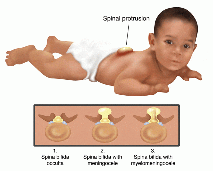
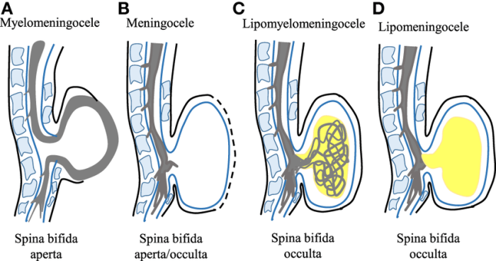
Image Description: A simple drawing of a spine with a small, barely visible gap in one of the vertebrae. No visible sac or bulge. Drawing Description: The spine is depicted as a series of stacked blocks representing vertebrae. One block shows a tiny, almost imperceptible gap. The surrounding tissue appears normal. Caption: Spina bifida occulta often shows no external signs. The defect is usually small and hidden under the skin. |
Image Description: A drawing of a spine with a visible sac protruding from the back. The sac is filled with fluid and is not directly connected to the spinal cord. Drawing Description: A small, fluid-filled sac (meningocele) is shown bulging from the back, at the site of a vertebral defect. The spinal cord is not directly involved in the sac. Caption: Meningocele involves a fluid-filled sac, but the spinal cord remains intact within the spinal canal. Okay, so you’re totally into drawing, right? Spina bifida easy drawings are a great way to learn anatomy, but sometimes you need a break. Check out these easy food labels drawing tutorials for a fun, chill change of pace. Then, get back to those awesome spina bifida drawings – you got this! |
Image Description: A drawing of a spine with a large, open sac protruding from the back. The sac contains both spinal fluid and a portion of the spinal cord. Drawing Description: A large, open sac (myelomeningocele) is shown, clearly protruding from the back. A portion of the spinal cord is visible within or attached to the sac. Caption: Myelomeningocele is the most severe form, with the spinal cord and its nerves exposed and potentially damaged. |
Anatomical Feature Comparisons
The key visual differences lie in the presence and size of the sac, and the involvement of the spinal cord. In spina bifida occulta, there’s minimal to no visible defect. Meningocele presents with a fluid-filled sac, but the spinal cord is typically unharmed. Myelomeningocele, however, shows a large, open sac containing portions of the spinal cord and nerves, leading to potentially significant neurological deficits.
The severity directly correlates with the extent of the spinal cord involvement and the resulting neurological damage. For instance, a child with myelomeningocele might experience paralysis or bowel and bladder dysfunction, whereas someone with spina bifida occulta may experience no symptoms at all.
Illustrating Associated Conditions

Spina bifida, sadly, often comes with other health challenges. Understanding these associated conditions is crucial for providing the best possible care and support. Think of it like this: it’s not just one thing, it’s a whole picture, and we need to see the whole picture to really help. These drawings will help visualize some of these common companions to spina bifida.Knowing what these associated conditions look like visually can help caregivers, doctors, and even the child themselves understand what’s going on and what to expect.
It’s all about empowering everyone involved.
Hydrocephalus
Hydrocephalus, a buildup of fluid in the brain, is a frequent complication of spina bifida. In a child’s drawing, hydrocephalus might be represented by a head that looks unusually large compared to the body. You might see the child drawing a head that’s swollen or rounder than normal. The eyes might be drawn slightly bulging. Another way a child might illustrate this is by drawing a wavy or swirling line inside the head, representing the excess fluid.
This isn’t a medical illustration, of course, but a child’s interpretation of what they might see or feel.
Chiari Malformation
Chiari malformation involves the cerebellum (the part of the brain that controls balance and coordination) extending down into the spinal canal. A child’s drawing might show a brain that seems to be “squeezed” or pushed down towards the neck area. They might draw a small, pinched area at the base of the skull or show the brain extending further down the spine than usual.
It’s a subtle visual, but the key is the altered shape and position of the brain stem within the skull.
Tethered Cord
Tethered cord syndrome happens when the spinal cord is attached too tightly to the surrounding tissues, hindering its growth and function. A child may not directly illustrate the cord itself being tethered, but might depict limited movement or unusual posture. For example, they might draw a child in a wheelchair, or a child with legs that are stiff or bent at an unusual angle.
The child may be drawing a leg that looks noticeably smaller or different than the other, indicating a possible nerve impairment.
- Hydrocephalus Drawing: A child draws a head significantly larger than the body, with slightly bulging eyes. The head might be shaded to show swelling.
- Chiari Malformation Drawing: A child draws a brain that appears compressed or pushed downwards towards the neck, with a smaller than normal brain stem.
- Tethered Cord Drawing: A child draws a child sitting in a wheelchair or with legs that are stiff and bent in an unusual way. One leg may be visibly smaller than the other.
Top FAQs
What are the potential limitations of simplified spina bifida drawings?
Simplified drawings, while helpful for understanding basic concepts, may omit crucial details relevant to specific cases. They should be used as educational tools and not as a substitute for professional medical advice.
How can I ensure my drawing is age-appropriate for children?
Use bright colors, simple shapes, and avoid complex medical jargon. Focus on the key features of the condition in a way that is easily understandable for the target age group. Consult with educators or child development specialists for guidance.
Where can I find resources for accurate anatomical references?
Medical textbooks, reputable online medical databases, and anatomical atlases are excellent resources for accurate anatomical references. However, remember to simplify the information for your intended audience.


